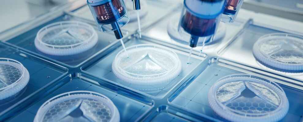
“In five to ten years, rather than just printing a model so we can run through a surgery beforehand, I foresee custom-made prostheses and custom-made cutting guides and other things that are very specific to streamline our surgery. I think 3D printing will become the norm for us, rather than only using it in special circumstances”
Stephanie Goldschmidt, DVM, Assistant Professor in Dentistry and Oral Surgery, University of California, Davis School of Veterinary Medicine
The advent of 3D printing technology has revolutionized medicine, offering unprecedented capabilities for creating customized medical solutions. Since its inception in the early 1980s, 3D printing has evolved from a rapid prototyping tool for industrial applications to a critical innovation in the medical field. Initially used to create dental implants and orthopedic devices, this technology has rapidly advanced to include the bioprinting of tissues and organs. Today, 3D bioprinting is making significant strides in veterinary care, facilitating personalized treatment plans and life-saving interventions.
Just like 3D printing is making waves in human medicine, it is revolutionizing veterinary care by providing novel solutions for complex medical challenges. Veterinarians have been using traditional 3D printing for some time to create custom prosthetics and orthopedic implants tailored to individual animals’ unique anatomy, thereby enhancing surgical interventions’ outcomes. 3D bioprinting takes this technology a step further by fabricating tissues and organs using living cells, which holds promise for regenerative therapies and personalized medicine in veterinary practice.
While traditional 3D printing is well-established in veterinary care, 3D bioprinting is still in its infancy. Despite its transformative potential, the widespread adoption of 3D bioprinting in veterinary care faces significant financial barriers. The high cost of 3D bioprinting technology, including the necessary equipment, materials, and specialized training, makes it prohibitively expensive for many practices and pet owners. Traditional 3D printing, on the other hand, is less expensive since it doesn’t require bioinks.
Dr. Stephanie Goldschmidt, an assistant professor in dentistry and oral surgery at the University of California, Davis School of Veterinary Medicine, has been using 3D printing in her practice for many years. Her primary use is to create 3D models of animals before surgery to help with the planning: “3D printing gives us the ability to better and more efficiently perform surgeries by allowing a tactile sensation of what we’re getting into, and that, right now for us, is the most beneficial,” she says.
Keep reading to learn more about the current and future applications of 3D printing in veterinary medicine.
Meet The Expert: Stephanie Goldschmidt, DVM, DAVDC

Dr. Stephanie Goldschmidt is a highly accomplished academic and an assistant professor in dentistry and oral surgery at the University of California, Davis School of Veterinary Medicine. Her primary focus is on surgical oncology and various oral maxillofacial surgeries.
Dr. Goldschmidt’s academic journey began with her undergraduate studies at the University of Wisconsin, Madison. She then pursued veterinary school at the University of Edinburgh in Scotland, where she earned her BVMS (bachelor of veterinary medicine and surgery). She then furthered her training through a rotating internship in New York and a residency at UW Madison. Notably, she is fellowship-trained in oral maxillofacial surgery. She is an accomplished researcher with numerous publications, including one in 2021 on using 3D dental models as an effective teaching tool. She is also a Diplomate of the American Veterinary Dental College (DAVDC).
Current Applications for 3D Printing in Veterinary Care
Traditional 3D printing has found extensive applications in veterinary care, fundamentally altering how veterinarians can approach complex medical procedures. By leveraging the capabilities of 3D printing, veterinarians can create highly accurate anatomical models, which are invaluable for surgical planning and training. These models facilitate a tactile understanding of an animal’s unique anatomy, allowing for precise preoperative assessments and more effective educational tools for students and practitioners alike.
“We will make 3D prints of complex trauma cases and tumors to help us explain exactly what needs to be done to the students and the owners. It also gives us a real tactile sense of what the surgery will be like,” shares Dr. Goldschmidt. “If we’re using any plates in the surgery we can also pre-contour all our plates, which makes our surgery more efficient and faster.”
While this technology is primarily used for all kinds of animals, it is particularly helpful for dogs with shortened or flattened muzzles. In the past, veterinarians have had to rely on 3D images to plan surgery: “The anatomy of the pet’s nose and respiratory apparatus can be very hard to visualize. This just gives us the best information as we go in to do these surgeries. The teeth can be especially problematic because there’s a lot of overlay. So even on a 3D CT scan, it can be difficult because the teeth are on top of teeth,” says Dr. Goldschmidt. With the 3D models in their hands, they can truly see what they will be working with during surgery.
3D printing is also used to fabricate custom prosthetics and orthopedic implants tailored to the specific needs of individual animals. This technology enhances the success rates of surgeries and improves postoperative recovery. It mitigates the risk of complications and ensures that interventions are more personalized and efficient, ultimately leading to better health outcomes for veterinary patients.
While Dr. Goldschmidt primarily works with traditional printing, 3D bioprinting is also slowly working its way into the veterinary field. As technology advances, there is potential for 3D bioprinting to facilitate regenerative medicine and personalized treatments for animals. This could include creating living tissues and organs using a pet’s own cells, eliminating the risk of rejection and the need for immunosuppressant drugs. It also allows for developing novel therapies and treatments that may not have been possible before.
However, the use of 3D bioprinting in veterinary medicine is still in its early stages, and further research and development are necessary before it becomes widely available.
Problems 3D Printing Solves In Veterinary Medicine
3D printing has been a game-changer in many areas of veterinary medicine, offering solutions to some long-standing problems. One of the primary benefits is improved surgical planning and accuracy. 3D printing creates highly accurate anatomical models, which can aid in preoperative assessments and surgical planning. This technology also enables the production of patient-specific surgical guides and instruments that improve the accuracy and efficiency of procedures: “It allows us to play through the surgery beforehand and ensure it’s more efficient. It also lets us know how thick parts of the surgery will feel,” says Dr. Goldschmidt.
Other areas where 3D printing excels in helping veterinary educators teach students anatomy and procedures. With 3D-printed models, students can better understand complex anatomical structures and surgical techniques before performing them on live animals. This allows for more practical and hands-on training. “When we show them a print, I think it helps them really understand what we’re talking about. We’re showing them a CT and running through the slices,” explains Dr. Goldschmidt.
3D printing technology also enables the creation of customized prosthetics and orthopedic implants tailored to an animal’s unique anatomy, thereby improving the success rates of surgeries and postoperative recovery. Additionally, it can help reduce costs associated with traditional surgical procedures that often require expensive equipment or materials by streamlining the processes and minimizing the time spent in surgery.
The Future of 3D Printing for Veterinarians
As the use of 3D printing in veterinary care continues to expand, there are exciting possibilities for its future applications. One area with potential is the development of customized medical devices and implants specifically designed for individual animals. This could include prosthetics, orthopedic implants, and dental appliances tailored to a pet’s needs.
Another area in which Dr. Goldschmidt sees potential growth is cutting guides. Cutting guides are precision-engineered tools used in veterinary surgery to enhance the accuracy of bone cuts. These guides are typically created using 3D printing technology, which allows for the customization of each guide to fit the unique anatomy of the individual animal.
By employing cutting guides, veterinarians can ensure that the bone is cut at the exact angle and depth required for effective surgical outcomes: “Our orthopedic service already is printing many cutting guides for their surgeries. I think that’s going to become more and more common in vet med as 3D prints become more and more accessible and not just at these tertiary care facilities,” she says.
However, a hurdle that must be overcome is the financial expense of these technologies: “Definitely, there is a little bit of a socioeconomic divide due to the cost of the print. Luckily, because we’re a university, we can keep that cost low,” Dr. Goldschmidt explains. “We decide to use it when it’s a really challenging case, and we build it into the cost of that surgery.”
As technology continues to improve and become more accessible, it is possible that these costs will decrease, making 3D printing a more widely available tool in veterinary medicine.
As with most new technologies, more infrastructure is needed to make them scalable. According to Dr. Goldschmidt, the bottleneck in leveraging 3D printing technology in veterinary medicine lies in transforming CT scan data into a usable STL file. CT scans provide intricate and detailed images of an animal’s anatomy, capturing nuanced structures and complex geometries.
However, converting these volumetric images into a format suitable for 3D printing is challenging and time-consuming: “Some clinicians will do it themselves if they’re familiar with the program. Most outsource it. We outsource that to our biomedical engineering department at the University. A commercial entity that is at a low enough cost that it could be outsourced to create the prints would make this all much more feasible,” she says.
“In five to ten years, rather than just printing a model so we can run through a surgery beforehand, I foresee custom-made prostheses and custom-made cutting guides and other things that are very specific to streamline our surgery. I think 3D printing will become the norm for us, rather than only using it in special circumstances,” Goldschmidt predicts.

